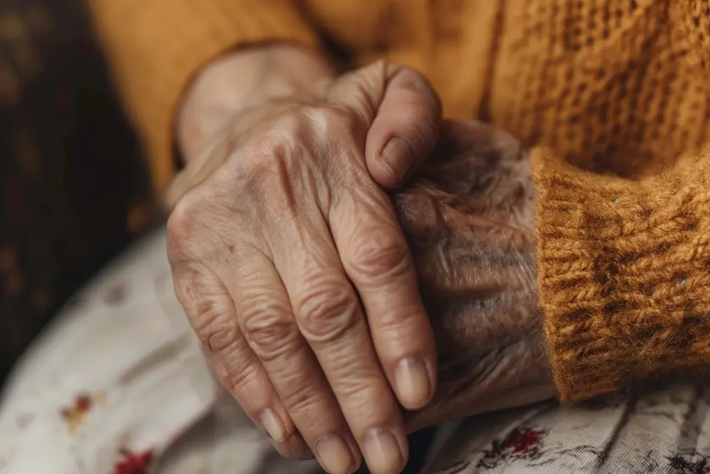
In a study of 500 elderly Indians attending a dermatology outpatient department, pruritus was the most common skin complaint, affecting 49.6% of individuals and in nearly 30% of cases, it was directly caused by xerosis, or pathologically dry skin. Wrinkles and age spots attract the most family attention. But it is the dry, itchy, thinning skin and the systemic diseases hiding behind it that carry the real clinical weight. Knowing the difference between what is expected in aged skin and what requires a doctor's attention is one of the most practically useful things a family caring for an elderly parent can learn.
Cosmetic Concern Versus Clinical Signal
When the family of a 72-year-old woman notices she is scratching constantly, the first response is usually a moisturiser. When they notice new dark spots appearing on her arms, they worry about sun damage and reach for a fairness cream. When they see her skin bruising easily or healing slowly after a minor scrape, they attribute it to "thin skin with age."
All three of these responses are understandable. All three miss opportunities for clinical investigation.
Persistent pruritus without a visible rash in an elderly person is not simply dry skin until it is proven to be dry skin. In the absence of skin lesions, underlying systemic illnesses, medications, nutritional status, occult malignancy, and psychological factors should be carefully evaluated in an elderly patient presenting with pruritus. Thyroid disease, chronic kidney disease, liver disease, iron deficiency anaemia, lymphoma, and certain medications are all documented systemic causes of generalised itch in older adults and they require blood tests, not moisturiser, to exclude.
The central challenge of skin ageing in elderly patients is precisely this: distinguishing between normal and abnormal changes, as well as physiological versus pathological alterations, is one of the primary clinical challenges in assessing geriatric skin. This blog is an attempt to draw that line clearly.
Two Distinct Ageing Processes, One Skin
Skin ageing in elderly patients is driven by two biologically distinct processes that operate simultaneously but through different mechanisms.
Intrinsic ageing also called chronological ageing is the genetically programmed, time-dependent degradation of skin structure that begins in the mid-twenties and accelerates significantly after 60. Both intrinsic and extrinsic ageing processes are associated with phenotypic changes in cutaneous cells, but the major functional manifestations of ageing occur as a consequence of structural and compositional remodelling of normally long-lived dermal extracellular matrix proteins. The key proteins affected are collagen which provides tensile strength and elastin, which gives skin its recoil and resilience.
As dermal fibroblasts age, they produce decreasing amounts of collagen and elastin. Simultaneously, they produce increased amounts of enzymes called matrix metalloproteinases (MMPs) that actively degrade these structural proteins. This dual process reduced production and increased degradation results in a drastic loss of collagen and elastin over time, manifesting clinically as skin laxity, fragility, fine wrinkling, and loss of the padding effect of subcutaneous fat.
The epidermis also thins, and the epidermal-dermal junction flattens a change with a specific clinical consequence: the thinned epidermis and flattening of the epidermal-dermal junction increase the incidence of skin tears from shearing and friction. This is why an elderly person bruises from a contact that would not mark a younger person's skin, and why their skin splits rather than stretches when caught on a sharp corner.
Extrinsic ageing predominantly driven by cumulative ultraviolet radiation exposure operates through an additional, overlapping mechanism. UV radiation generates reactive oxygen species that damage collagen and elastin fibres independently of the intrinsic pathway. Long-lived proteins in the dermal matrix and cytoskeleton are particularly susceptible to glycation, resulting in tissue stiffening and reduced elasticity and glycated elastin fibres abnormally aggregate, contributing to the characteristic rough, thickened, deeply wrinkled appearance of photoaged skin, distinct from the finer wrinkling of pure chronological ageing.
In Indian skin predominantly Fitzpatrick type IV and V an important finding from Indian dermatological studies provides important context: photoageing changes were less common than chronological ageing changes in skin type IV, with 83% of cases showing chronological ageing without photoageing. This means that for most urban Gurgaon seniors with darker skin, the primary driver of visible skin ageing is intrinsic and time-dependent not solar damage though UV protection remains clinically important for malignancy prevention.
The Clinical Reality for Indian Seniors in Gurgaon
Indian dermatological studies from tertiary hospitals provide a clear picture of what elderly Indian skin actually presents with and it differs from what Western literature emphasises.
Xerosis was noted in 88.5% of elderly patients in a North Indian study, and pruritus was the commonest complaint at 80%. These two conditions dry skin and itching are almost universal in Indian elderly patients and are frequently undertreated. Xerosis is not merely uncomfortable: untreated xerosis can progress to eczema craquelé a cracked, fissured eczematous condition and increases susceptibility to stasis dermatitis and ulcer formation.
Seborrhoeic keratosis, a benign warty overgrowth, was the most common benign neoplasm found in elderly Indians, affecting 50.6% of study participants a finding that causes considerable family anxiety when discovered, since the lesions can look alarmingly like malignant growths to non-medical observers. Seborrhoeic keratoses are not pre-malignant and do not require treatment unless they cause discomfort or bleed repeatedly.
The systemic disease connection is well established in Indian data. Diabetes mellitus often manifests in skin as xerosis, pruritus, or diabetic dermopathy, while chronic renal disease may present with pallor, pruritus, or calciphylaxis. Cardiovascular disease can cause stasis dermatitis and leg ulcers due to compromised vascular efficiency. For urban Gurgaon seniors managing multiple chronic conditions as most do a new or worsening skin symptom is therefore frequently a systemic signal deserving investigation, not a standalone dermatological problem.
The air quality dimension is locally relevant. Exposure to pollutants can trigger inflammatory skin disorders such as eczema or psoriasis and Gurgaon's seasonal pollution spikes, combined with the dry indoor air created by year-round air conditioning, create a compounded drying and irritant environment for elderly skin that is already compromised by intrinsic ageing.
What Is Normal, What Is Not: A Clinical Distinction Guide
Expected and not requiring urgent investigation:
Fine wrinkles and skin laxity; loss of subcutaneous volume in the face and hands; easy bruising from minor contact; slower wound healing; scattered seborrhoeic keratoses (stuck-on, waxy, brown-black lesions with a rough surface); senile lentigines (flat, evenly pigmented brown spots in sun-exposed areas); skin tags (acrochordons) in body folds; and mild, diffuse dry skin responsive to moisturiser.
Requires dermatological evaluation:
Any skin lesion that is growing, bleeding, changing colour, or developing an irregular border; a non-healing wound or ulcer present for more than two weeks; new widespread rash appearing alongside a new medication; persistent generalised itch without visible rash, or itch unresponsive to moisturisers after two weeks of consistent use; sudden onset of multiple seborrhoeic keratoses (the sign of Leser-Trélat rapid appearance of multiple keratoses can rarely indicate internal malignancy); skin thickening and darkening in the armpits or groin (acanthosis nigricans associated with insulin resistance); and any purplish, non-blanching spots on the legs.
Practical Steps: What Families Can Do
1. Treat xerosis as a medical condition, not a cosmetic problem
Daily moisturisation with an emollient applied immediately after bathing while skin is still slightly damp, to lock in moisture is the most impactful intervention for xerosis in elderly patients. Emollients with additional antipruritic agents such as menthol or camphor can temporarily reduce pruritus alongside addressing the dryness. Avoid fragrance in moisturisers fragrance is a common contact irritant in aged skin. Soap should be replaced with a soap-free, pH-balanced cleanser. Bathing in hot water worsens xerosis; use lukewarm water only.
2. Do not accept persistent itch as simply "dry skin" without investigation
If generalised itch persists despite two weeks of consistent emollient use, request a blood panel that includes CBC, kidney function, liver function, thyroid function, and fasting glucose before attributing the itch to xerosis alone. Pruritus without a visible rash in an elderly patient is a systemic search question, not a dermatology-first question.
3. Learn to distinguish seborrhoeic keratoses from melanoma
Seborrhoeic keratoses have a characteristic appearance: stuck-on, with a rough, verrucous surface, well-defined borders, and a uniform colour that ranges from light brown to black. Melanoma, by contrast, shows the ABCDE features: Asymmetry, Border irregularity, Colour variation within the lesion, Diameter over 6mm, and Evolution over time. Any lesion the family is uncertain about should be assessed by a dermatologist not monitored at home.
4. Protect from UV exposure not for cosmetic reason but for malignancy prevention
Even in darker Indian skin types with lower baseline photoageing, cumulative UV exposure is a documented risk factor for basal cell carcinoma and squamous cell carcinoma in elderly patients. A broad-spectrum SPF 30 or higher applied to sun-exposed areas face, forearms, the back of the hands on a daily basis is a cancer prevention measure, not a cosmetic preference. This is particularly relevant for Gurgaon seniors who walk outdoors in the morning.
5. Check medications as a cause of skin changes
Many medications commonly prescribed to elderly patients cause or worsen skin symptoms: anticoagulants cause easy bruising; diuretics worsen xerosis; calcium channel blockers cause ankle oedema and skin changes; certain antibiotics and NSAIDs cause photosensitivity rashes; and long-term steroids cause skin thinning and fragility. If a skin change appeared or worsened after a new medication was started, this connection must be raised with the prescribing physician.
At Aamra Seniors Club, clinical observation is built into our day programme which means subtle skin changes, wounds, and systemic signals do not go unnoticed between annual check-ups. Book a Day Pass.
Critical Warning: Any wound, ulcer, or skin break on the leg or foot of an elderly patient with diabetes or peripheral vascular disease that has not healed within two weeks requires urgent medical assessment not home dressing changes and observation. Diabetic foot ulcers and venous leg ulcers can deteriorate rapidly and silently in elderly patients with reduced pain sensation. Additionally: any skin lesion that bleeds spontaneously, grows visibly over weeks, or has an irregular border should be assessed by a dermatologist within two to four weeks, not at the next convenient appointment.